Researchers in the United States and Spain have discovered that a tool widely used in nanoscale imaging works differently in watery environments, a step toward better using the instrument to study biological molecules and structures.
The researchers demonstrated their new understanding of how the instrument – the atomic force microscope – works in water to show detailed properties of a bacterial membrane and a virus called Phi29, said Arvind Raman, a Purdue professor of mechanical engineering. An atomic force microscope uses a tiny vibrating probe to yield information about materials and surfaces on the scale of nanometers, or billionths of a meter. Because the instrument enables scientists to “see” objects far smaller than possible using light microscopes, it could be ideal for studying molecules, cell membranes and other biological structures. The best way to study such structures is in their wet, natural environments. However, the researchers have now discovered that in some respects the vibrating probe’s tip behaves the opposite in water as it does in air, said Purdue mechanical engineering doctoral student John Melcher. The probe is caused to oscillate by a vibrating source at its base. However, the tip of the probe oscillates slightly out of synch with the oscillations at the base. This difference in oscillation is referred to as a “phase contrast,” and the tip is said to be out of phase with the base.
Although these differences in phase contrast reveal information about the composition of the material being studied, data can’t be properly interpreted unless researchers understand precisely how the phase changes in water as well as in air, Raman said.
If the instrument is operating in air, the tip’s phase lags slightly when interacting with a viscous material and advances slightly when scanning over a hard surface. Now researchers have learned the tip operates in the opposite manner when used in water: it lags while passing over a hard object and advances when scanning the gelatinous surface of a biological membrane.
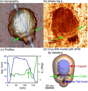
Researchers deposited the membrane and viruses on a sheet of mica. Tests showed the differing properties of the inner and outer sides of the membrane and details about the latticelike protein structure of the membrane. Findings also showed the different properties of the balloonlike head, stiff collar and hollow tail of the Phi29 virus, called a bacteriophage because it infects bacteria.
Original Publication:
Melcher J, Carrasco C, Xu X, Carrascosa JL, Gómez-Herrero J, José de Pablo P, Raman A. (2009): Origins of phase contrast in the atomic force microscope in liquids. Proc Natl Acad Sci U S A. 2009 Aug 18;106(33):13655-60. Epub 2009 Aug 5.

Researchers in the United States and Spain have discovered that an atomic force microscope - a tool widely used in nanoscale imaging - works differently in watery environments, a step toward better using the instrument to study biological molecules and structures. The researchers demonstrated their new understanding of how the instrument works in water to show details of the mechanical properties of a virus called Phi29. The images in "a" and "c" show the topography, and the image in "b" shows the different stiffness properties of the balloonlike head, stiff collar and hollow tail of the Phi29 virus, called a bacteriophage because it infects bacteria. (C. Carrasco-Pulido, P. J. de Pablo, J. Gomez-Herrero, Universidad Autonoma de Madrid, Spain)


